The Continuous Loop of Arteries at the Base of the Brain is Called
Linda Crampton is an experienced teacher with a first-class honors degree in biology. She writes about the scientific basis of disease.
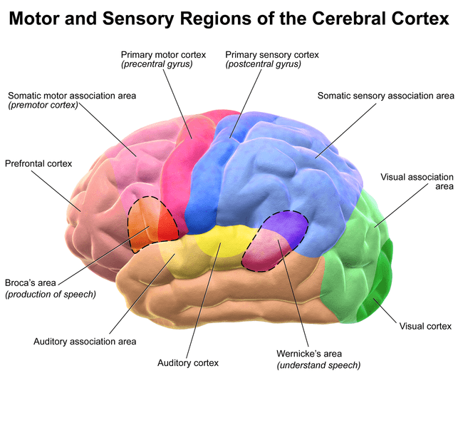
This illustration shows colour-coded lobes of the cerebral cortex. Pink = frontal lobe, blue = parietal lobe, orange = temporal lobe, green = occipital lobe
BruceBlaus, via Wikimedia Commons, CC BY 3.0 License
The Amazing Human Brain
The human brain is a fascinating and very complex organ that is only slowly giving up its secrets. It has taken us thousands of years to reach our current state of knowledge about the organ. We still don't understand everything about its structure and function. Many researchers are investigating the brain's activities, however, since it's such a vital part of our lives.
In the past, there was a tendency to name newly discovered body structures after their discoverer. This article describes three brain regions and also includes some facts about the physician-scientists who are forever (as far as we know) linked to them.
Broca's area was named after Paul Broca, a French doctor of the nineteenth century. Carl Wernicke was a German physician. He gave his name to Wernicke's area and lived until the start of the twentieth century. The circle of Willis was named after Thomas Willis, an English doctor from the seventeenth century.
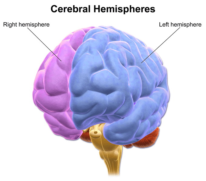
The cerebral hemispheres as viewed from the front of the brain
BruceBlaus, via Wikimedia Commons, CC BY 3.0 License
An Overview of Brain Organization
The cerebrum is the largest and most obvious part of the brain. It consists of two halves, which are known as cerebral hemispheres. The hemispheres are joined together by three commissures. The biggest one is a band of tissue called the corpus callosum. The anterior and posterior commissures are smaller.
Although this article refers to processes that occur in the cerebral hemispheres, the sections underneath the structures are also important in our lives. The so-called higher functions of the brain, such as thinking, reasoning, making decisions, and speaking, originate in the cerebrum, however.
Each hemisphere of the cerebrum consists of four visible lobes known as the frontal, parietal, temporal, and occipital lobes. Broca's area is a patch of tissue located in one of the two frontal lobes. It's usually found in the left hemisphere, as shown below, but it's sometimes located in the right one.
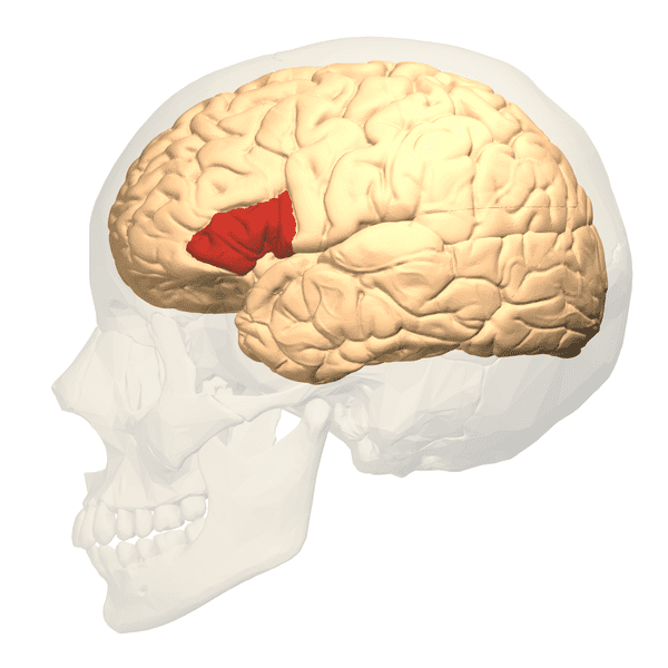
Broca's area (the red patch) is located in a frontal lobe of the cerebrum.
Database Center for Life Science, via Wikimedia Commons, CC BY-SA 2.1 JP
Facts About Broca's Area
Broca's area plays an important role in the creation of speech. People who have damage in this area have great difficulty in speaking, even when there is nothing wrong with the rest of their brain or with the body's mechanical components for forming spoken words. Patients may be able to speak a few to a significant number of words but can generally create only short sentences. They often need to pause as they speak. The reasoning and thinking parts of their brain are usually unaffected, so the situation can be very frustrating for them. The disorder is commonly known as Broca's aphasia. It's also referred to as expressive or non-fluent aphasia.
Aphasia is an impairment in the ability to produce or understand speech or written material. The disorder ranges from a mild condition to a severe one. Modern treatment techniques (and in some cases the body's healing mechanisms after an injury) may help aphasia. The teenager in the video below is recovering from a stroke and Broca's aphasia.
Paul Broca Information
Broca's area was discovered by a French neurosurgeon named Paul Broca (1824–1880). In 1861, Broca examined the brain of a man who had recently died. Although the man had been able to produce sounds, the only recognizable word that he had been able to say was "tan". Broca discovered a damaged area in the man's left frontal lobe. He subsequently found damage in the same brain area in other people with similar speech problems. Broca concluded that he had found the part of the brain that was responsible for speech.
Two of the brains that Broca examined were preserved, including the brain of his first patient. Both patients had been severely limited in their speech. In 2007, scientists performed an MRI scan of the preserved brains. They found that although in each case Broca's area was damaged, the injury extended further into the brain. The extent of an injury as well as its exact location likely contribute to the problems experienced by someone with Broca's aphasia.
Broca's area isn't directly responsible for speech. It sends nerve signals to the motor cortex, which stimulates the muscles in the mouth and face to contract in order to produce words.
Scroll to Continue
Read More From Owlcation
Marc Dax (1770–1837) was a French neurologist who discovered that damage to a region of the left hemisphere of the brain can cause speech problems. Since he did this before Broca's research, some people think that both scientists should share the honour of the discovery.
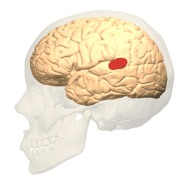
Wernicke's area is located where the parietal lobe joins the temporal lobe.
Database Center for Life Science, via Wikimedia Commons, CC BY-SA 2.1 JP
Wernicke's Area Facts
About ten years after Broca's discovery, a scientist named Carl Wernicke discovered another area that is often located on the left side of the brain and is related to speech. Wernicke's area is located mostly in the temporal lobe and partially in the parietal lobe. It's involved in understanding the meaning of spoken words.
People with damage to Wernicke's area can often speak fluently, but what they say makes no sense in relation to the situation. They may sometimes use made-up words as well as real ones and may not show any awareness that they've done this. In addition, they sometimes have a tendency to speak excessively. The disorder is known as Wernicke's aphasia. It's also referred to as receptive or fluent aphasia. Patients may have trouble with understanding written language as well as speech.
Broca's area and Wernicke's area are connected by a bundle of nerve fibres, forming what is known as a language loop. Both areas are important in producing intelligible speech.
This language loop is found in the left hemisphere in about 90% of right-handed persons and 70% of left-handed persons, language being one of the functions that is performed asymmetrically in the brain.
— The Brain page from McGill University
Some researchers are re-examining the functions of Broca's and Wernicke's areas. Though they are language-related areas, their functions may not be exactly what their discoverers believed. Even some modern assumptions about their jobs may be incorrect. The brain is complex and a difficult organ to study.
Information About Carl Wernicke
Carl Wernicke was a German doctor who was born in 1848. He was killed in an accident in 1905, reportedly while riding his bike. Wernicke is often classified as a neuropsychiatrist. He believed that patients with psychiatric problems had problems in a specific region or pathway in their brain rather than in the brain as a whole.
Wernicke discovered the region now named in his honour and found that damage in the area produced aphasia. He was only 26 years old when he published the results of his discovery. He referred to the disorder resulting from the damage as sensory aphasia. The name was later changed to honour his work.
Wernicke is also known for other publications, including a three-volume work created between 1881 and 1883. The work was called Textbook of Brain Disorders. In the publication, Wernicke linked different neurological problems to specific areas of the brain. It's a shame that he died at a comparatively young age.
Aphasia is often caused by strokes, especially in older people. Strokes occur in younger people as well, however. Aphasia may also develop due to tumours, head trauma, and infections.
The Circle of Willis
The circle of Willis is a roughly circular network of arteries located on the undersurface of the brain. Although it belongs to the circulatory system instead of the nervous system, it's often referred to as part of the brain. The arteries play a role in the circulation of blood through the brain.
The circle of Willis is an example of a circulatory anastomosis—a structure in which there is a cross-connection between blood vessels that we would expect to stay separate, such as two different arteries. Normally, blood circulates through the heart and blood vessels in the following sequence: the heart, an artery, arterioles, capillaries, venules, a vein, the heart. In the circle of Willis, blood flows from one artery to another one.
An anastomosis can provide a backup route for blood if the main passageway is blocked. As can be seen in the illustration below, arteries are connected to one another in the circle of Willis. Interestingly, many people have an atypical circle of Willis. Nevertheless, it's thought that the alternate blood route that it provides can be very useful in certain disorders.
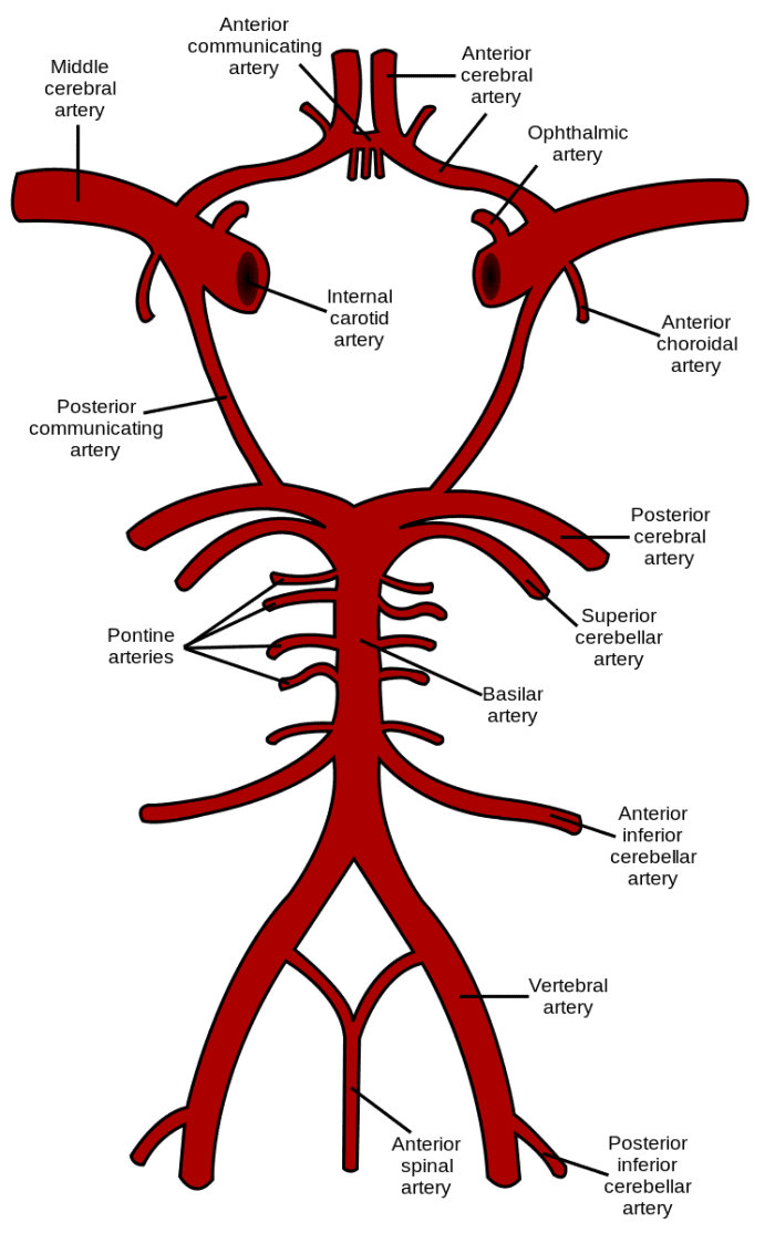
Most of the arteries in this illustration are present in a right and a left form. Only one of each pair is labelled. The circle of Willis is the roughly circular section at the top of the illustration.
Rhcastilhos, via Wikimedia Commons, public domain license
Components of the Circle of Willis
The arteries that make up the circle of Willis are divided into an anterior group (located nearer to the front of the brain) and a posterior group (located nearer to the back of the brain). The orientation of the circle is shown in the illustration below.
In order to make the illustrations above and below easier to understand, the arteries are cut off at the ends where they disappear from view, change direction, or are no longer considered to be part of the circle of Willis. The blood vessels that make up the circulatory system are actually continuous. They branch and merge and change in diameter and direction, but they never simply end.
The anterior group of arteries in the circle of Willis consists of the following blood vessels.
- Right and left anterior cerebral artery
- Anterior communicating artery (which is not paired)
- Right and left internal carotid artery
The posterior group consists of these vessels.
- Right and left posterior communicating artery
- The horizontal parts of the right and left posterior cerebral arteries
- The tip of the basilar artery (which is not paired)
Blood is sent from the heart to the brain (and to the circle of Willis) through the two internal carotid arteries and the two vertebral arteries.
Thomas Willis Facts
Thomas Willis was an English physician who was born in 1621 and died in 1675. He is often said to be the father of neurology. Neurology is the study of the nervous system.
Since the blood vessels at the base of the brain are visible to the unaided eye, other people noticed the circle of arteries before Willis did. Willis is credited with the circle's discovery, however, due to his meticulous and detailed observations that were far superior to previous attempts to describe the region.
Willis's discoveries were published along with other brain observations in 1664 in a book entitled Cerebri Anatome. The title is a Latin term meaning Anatomy of the Brain. At the time when Willis was alive, scientists created their publications in Latin. Christopher Wren created the illustrations for Cerebri Anatome. He is famous today for his design of St. Paul's Cathedral in London.
The Importance of Brain Research
The brain still holds many mysteries. In July 2016, scientists working on the Human Connectome Project in the United States announced that they had discovered 97 new regions of the brain. The project is run by the National Institutes of Health (NIH) in the United States. Its goal is to map the neural pathways in the brain. The plan is very ambitious but has enormous implications in the realm of health and disease.
In the future, scientists may discover new facts related to the function of Broca's and Wernicke's areas and the circle of Willis. This will not only be interesting biologically but may also be useful in helping people recover from brain damage. Discovering more information about how the brain works is fascinating and potentially very important. It's a complex but vital organ in our lives.
References
- "Broca's Area, Wenicke's Area, and Other Language-Processing Areas in the Brain" from McGill University
- The Brain and Language from the University of Washington
- Circle of Willis anatomy from Radiopaedia
- Facts about the Circle of Willis from Medscape (Overview only)
- Paul Broca facts from the New World Encyclopedia
- Carl Wernicke information from Judy Duchan, University at Buffalo (State University of New York)
- Information about Wernicke from Encyclopedia.com
- Thomas Willis and Cerebri Anatome from the Journal of the Royal Society of Medicine
This content is accurate and true to the best of the author's knowledge and does not substitute for diagnosis, prognosis, treatment, prescription, and/or dietary advice from a licensed health professional. Drugs, supplements, and natural remedies may have dangerous side effects. If pregnant or nursing, consult with a qualified provider on an individual basis. Seek immediate help if you are experiencing a medical emergency.
© 2016 Linda Crampton
Linda Crampton (author) from British Columbia, Canada on September 22, 2019:
I'm very sorry about your brother-in-law's situation. I can't help you, though. I'm a science writer, not a doctor. Even a doctor would have to be familiar with a patient's specific case in order to answer your questions. I hope you find a physician that is able to answer the questions and help your brother-in-law.
Florence Njoku on September 22, 2019:
I have a brother in-law who had severe brain injury last three years and lost his power of speech since and quadriplegic too although the spinal cord is intact.
I am wondering could it be that his Broca's area was damaged as a result of the injury. He understands spoken word and has a strong grasping reflex. I consulted the best neurosurgeon at the time of his injury and was told nothing could be done for him. The hospital he was admitted severe brain damage and was querying brain death, I guess if if was brain death who will be dead and would not be able to breath or last weeks without life-support.
I am just wondering what could be the possible cause of him not being able to respond to treatment or to the natural power of the body to heal itself?
Linda Crampton (author) from British Columbia, Canada on August 06, 2019:
Hi, Bikash. Scientists gain knowledge from experiments with animal brains and from some types of human brain surgery where the patient is conscious. The person feels no pain, but they are kept conscious so the surgeon doing the brain surgery can make observations or get feedback about certain procedures.
Bikash Ghosh on August 02, 2019:
How the scientists know that the cerebrum memorize our information and processing our work?
Linda Crampton (author) from British Columbia, Canada on March 31, 2019:
Thank you for the comment. I've been doing some searching and discovered that some researchers do say that Broca's and perhaps Wernicke's area may be involved in some cases of autism. At this stage, it seems to be only a possibility, but the research is interesting.
Kathryn Gardner on March 25, 2019:
I thoroughly enjoyed this article. My question has to do with nonverbal, autistic children. Has there been research done to see if there is damage to any of these parts of the brain in autistic children? If so, is there any kind of stimulation that can be done in order for the child to speak?
Linda Crampton (author) from British Columbia, Canada on August 08, 2017:
Thanks, Anita. There are lots of interesting things to discover here.
Anita Hasch from Port Elizabeth on August 08, 2017:
Very interesting and informative. I love reading articles on hubpages as I seem to be learning something new all the time.
Linda Crampton (author) from British Columbia, Canada on April 18, 2017:
Thank you very much for the kind comment, Nadine. The cause of consciousness fascinates me, especially since it's not well understood.
Nadine May from Cape Town, Western Cape, South Africa on April 18, 2017:
Wow what an informative article on such a mysterious subject.We know still so little about the human brain, not to mention the topic of consciousness. Love the images and video. Well done.
Linda Crampton (author) from British Columbia, Canada on September 05, 2016:
Thank you for the comment, Deb. I appreciate it very much. I agree with you - it's going to take time to understand the brain in more detail. It's a complicated and amazing organ.
Deb Hirt on September 05, 2016:
It will still take time to learn the functions of the complicated makeup of the brain, for it controls so much of our bodily functions. This was a wonderful and fascinating read.
Linda Crampton (author) from British Columbia, Canada on August 02, 2016:
Thanks, Dianna. I appreciate your visit and comment, as I always do.
Linda Crampton (author) from British Columbia, Canada on August 02, 2016:
Hi, Devika. Thank you for the kind comment.
Dianna Mendez on August 02, 2016:
I enjoyed your post on the brain. I always learn so much from your sharing.
DDE on August 02, 2016:
Hi a fascinating subject and you presented in detail. A must read hub indeed.
Linda Crampton (author) from British Columbia, Canada on August 01, 2016:
Thank you very much, MsDora. I appreciate your visit a great deal.
MsDora on August 01, 2016:
Alicia, thanks for this very informative lesson. I will save it as a reference article to read again.
Linda Crampton (author) from British Columbia, Canada on July 31, 2016:
Thank you for the lovely comment, Faith. I appreciate it very much! The human brain is definitely awesome. It has many very impressive and amazing abilities.
I hope you're having a good weekend and that the week ahead is enjoyable for you.
Faith Reaper from southern USA on July 31, 2016:
Fascinating and well-written article on the amazing brain, Linda! Nothing man can ever come up with to come close to imitating the brain, even the most advanced computer in the world, nothing compares to the brain. This would fit nicely in my series, Our Amazing Bodies ... and I know I couldn't have covered the brain as well as you have here, no doubt!
It is certainly amazing they are still discovering new areas of the brain.
As always, I learned a lot once again.
I hope you are enjoying a peaceful weekend.
Linda Crampton (author) from British Columbia, Canada on July 30, 2016:
Thank you, Manatita. I appreciate your kind comment and the interesting information about Bruce Lee. I like his idea!
manatita44 on July 30, 2016:
An inspirational piece with very well-researched and sound knowledge. I was reading a little about Bruce Lee today. He feels that nothing is perfect, that there is and will always be scope for improvement. In this Light, I have no doubt that man will go further and yet further is this interesting study of the anatomy and indeed himself. Excellent Alicia.
Linda Crampton (author) from British Columbia, Canada on July 29, 2016:
Thank you very much, Larry. I appreciate your comment.
Larry Rankin from Oklahoma on July 29, 2016:
Really interesting science history. Wonderfully informative.
Linda Crampton (author) from British Columbia, Canada on July 29, 2016:
Hi, Bill. I appreciate your visit and comment, as always. I think that science is a very interesting topic.
Linda Crampton (author) from British Columbia, Canada on July 29, 2016:
Thank you very much for the interesting comment, Buildreps. I remember reading about the research that you describe. It certainly is fascinating! The activity of the brain is so intriguing.
Bill Holland from Olympia, WA on July 29, 2016:
What's really amazing is that many Americans don't use theirs. LOL
Great science lesson, as always. I love this stuff, so thank you!
Buildreps from Europe on July 29, 2016:
Interesting research and your article is fascinating. There is still so much to discover about the brain. A few years ago I watched a scientific series about the brain (don't know the name anymore; my brain is letting me down..), and what specifically fascinated me was a research where they attached sensors on the "subject's" head, and asked this person all kinds of questions (and tests etc). It appeared that the sensors/computer already picked up activity in the specific part that was expected to solve that specific question, but the person was not yet aware of the fact that he/she already solved the question. The research suggested that the unconscious part of the brain already solved it, but that it yet had to boil up to the conscious brain. Fascinating stuff. Not sure whether my comment is completely on topic though. Thanks for the great article.
Linda Crampton (author) from British Columbia, Canada on July 28, 2016:
Hi, Bill. Thank you very much for the comment. I agree - the brain is definitely amazing. It's a very interesting organ to study.
Bill De Giulio from Massachusetts on July 28, 2016:
Fascinating Linda. As always, thank you for the education. The brain is an amazing organ. As much as we know about it there seems to be much that we have yet to learn. Thank you for explaining things in simple, easy to understand language, which you do exceedingly well.
Linda Crampton (author) from British Columbia, Canada on July 28, 2016:
Thank you very much, Mel. I appreciate your comment. I like your description of the brain!
Mel Carriere from Snowbound and down in Northern Colorado on July 28, 2016:
Fascinating study about the language centers of the brain. The brain is more vast of an unexplored area than the cosmos, I think. Fantastic research!
Linda Crampton (author) from British Columbia, Canada on July 28, 2016:
Thank you for the visit and the comment, Flourish. I find it very interesting that scientists are still learning more about the brain, too. It's an awesome organ!
FlourishAnyway from USA on July 28, 2016:
I recall learning about the first two in college but not the Circles of Willis. Thanks for giving some of the biographical information behind the doctors. Well written and interesting. I find it fascinating that they are still discovering many specify parts of the brain and what they do.
Source: https://owlcation.com/stem/Exploring-the-Brain-Three-Regions-Named-after-Scientists
0 Response to "The Continuous Loop of Arteries at the Base of the Brain is Called"
Post a Comment